


Click on titles to open relevant information.
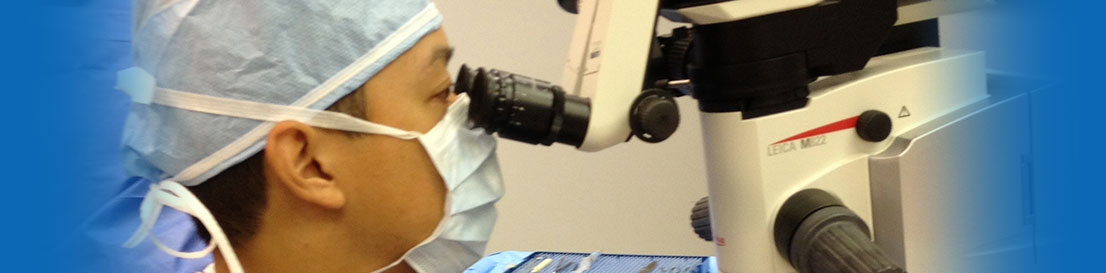
CATARACT SURGERY
Cataract surgery is a minimally invasive procedure performed on an outpatient basis at Naples Premier Surgery Center. The eye is anesthetized with topical and/or local anesthesia along with IV sedation. A small incision is made, into which an ultrasound probe is inserted to break up or emulsify the cataract into tiny pieces which are then suctioned out of the eye. Once the cataract has been removed, a new artificial intraocular lens is inserted to help improve vision. A stitch is usually not even needed!
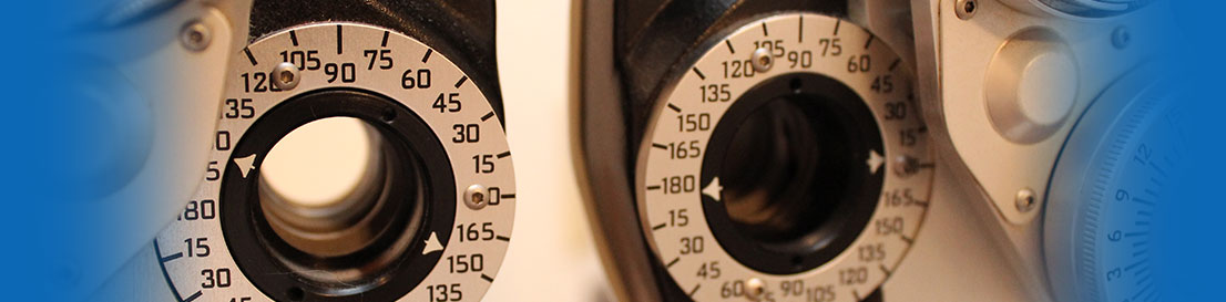
Each surgical patient is meticulously tested prior to cataract surgery to determine which intraocular lens is best for them. Dr. Tran and Dr. Vu employs the newest technology in intraocular lenses, including the Acrysof® Toric lens for astigmatism, the Restor® multifocal lens, the Tecnis multifocal lens, and the Crystalens® accommodative lens. Each of these lenses offers different advantages for post-surgical vision. The most effective lens depends on each patient’s individual preferences and goals for their vision.
RESTOR made by Alcon

Tecnis Multifocal by AMO

CRYSTALENS made by Bausch and Lomb

Toric Intraocular Lens
Toric IOLs are specifically designed for patients with astigmatism. In the past, patients with astigmatism would need eyeglasses or contact lenses even after cataract surgery. Toric IOLs such as the Acrysof® Toric corrects cataracts and astigmatism with just one lens, providing a convenient and affordable solution to your vision needs.
Multifocal Intraocular Lens
In the past, intraocular lenses were monofocal, meaning they were only able to correct distance vision, often leaving patients with the need for reading glasses. Multifocal IOLs such as the Tecnis multifocal or Restor® offer patients freedom from glasses after cataract surgery by improving vision at all distances. Up to 80% of patients do not need to rely on glasses with the Restor® multifocal IOL.
For more information please visit acrysofrestor.com
Accommodative Intraocular Lens
Accommodative IOLs such as the Crystalens® offer a full range of restored vision for cataract patients. The Crystalens® mimics the natural lens’ accommodative capabilities, allowing the patient to see clearly across a wide range of distances.
For more information please visit crystalens.com
Laser Cataract Surgery
Laser-based cataract surgery is when a femtosecond laser is used to break the eye’s natural lens into fragments for easier removal through an incision. A laser may also be used to make incisions into the eye during the cataract procedure.
Naples Premier Surgery Center is one of very few ophthalmology practices in the country to have access to this technology and your surgeon will determine whether or not laser cataract surgery should be included in your treatment plan.
ADVANCED LASER CATARACT SURGERY
Naples Premier Surgery Center now offers the LENSAR Laser System as part of your advanced cataract procedure because it is safe, effective and uses the same proven laser technology that’s been used in LASIK procedures for over a decade.
What is LENSAR and how does it improve my cataract procedure?
Your eye works a lot like a camera, using a lens to focus on an image. If your camera lens became cloudy, you'd have a hard time viewing the world around you. Just like a camera, the lenses in your eyes can become cloudy as you age, making it harder for you to see. This natural condition, known as a cataract, affects more than half of Americans by age 80.
The LENSAR Laser System allows your surgeon to offer you a better,more precise cataract removal procedure that is customized to your eye. With the LENSAR Laser System, your surgeon can remove your cataract in a more advanced way. Using the LENSAR Laser System ensures that your customized cataract procedure is performed with laser precision. This is because of Augmented Reality, a unique imaging system that provides your surgeon with a reconstructed 3-D view of your eye, in order to help plan and treat your cataract.
Want to Learn More? Click Here
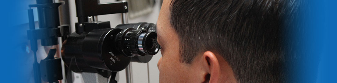
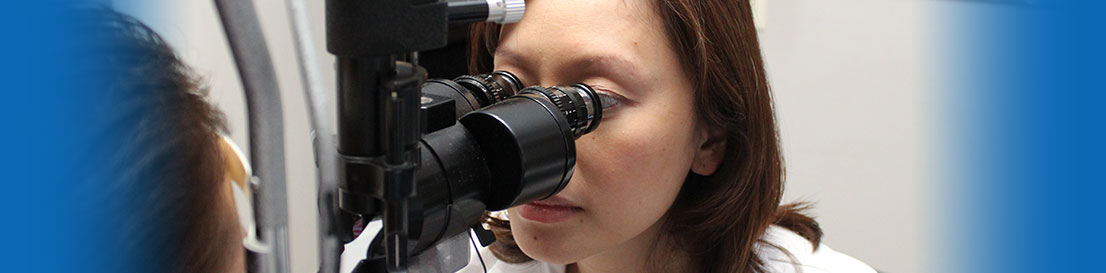
ORA- Optiwave Refractive Analysis
It used to be that your surgeon needed to wait weeks after performing surgery to determine your vision results. Thanks to ORA, this is no longer the case. Now, during the procedure, your surgeon can take measurements of your eye, make any necessary adjustments and refine your visual outcome. ORA measurements are taken after the clouded cataract is removed, when the surgeon has a clear view, allowing your surgeon to tailor your procedure to your individual eye. No matter what premium cataract procedure you and your surgeon decide upon, you can feel confident that by adding the ORA System, you’ll receive the best possible results.
GLAUCOMA SURGERY AND LASERS
Lasers
Selective Laser Trabeculoplasty (SLT)
SLT is a laser that treats the drain directly to help increase the outflow of fluid. It treats specific cells "selectively," leaving the trabecular meshwork intact. For this reason, SLT may be safely repeated. It is not painful, and often can be an alternative to eye drops in early open angle glaucoma.
Laser Peripheral Iridotomy (LPI)
Angle-closure glaucoma, also called closed-angle glaucoma, occurs when the iris bulges forward to narrow or block the drainage angle formed by the cornea and the iris. As a result, aqueous fluid can no longer reach the trabecular meshwork at the angle.
LPI creates a small hole in the iris, allowing it to fall away from the drainage angle and unblock the drain.
Surgeries
Trabeculectomy (Filtration Surgery)
During trabeculectomy (sometimes also called filtration surgery), a new drainage opening is created to bypass the clogged drainage channels of the trabecular meshwork. The opening is partially covered with a flap of tissue from the sclera (the white part of the eye) and the conjunctiva (the clear thin covering over the sclera). This new opening allows fluid (aqueous humor) to drain out of the eye under the conjunctiva and form a little blister, or bubble, called a bleb. The bleb is located just under the upper eyelid, where it is not visible.
Tube-Shunt Surgery for Glaucoma
Tube-shunt surgery involves placing a flexible plastic tube with an attached silicone drainage pouch in the eye to help drain fluid (aqueous humor) from the eye. This type of surgery is usually done after a failed trabeculectomy. If a person already has or is likely to form scar tissue in the eye, this type of surgery may be done initially. Tube-shunt surgery is outpatient, and is done under local anesthesia. A Baerveldt, Molteno, or Ahmed tube shunt may be used by your doctor, depending on the type of glaucoma, and the lowering of IOP needed.
Canaloplasty for Glaucoma
One recent state-of-the-art surgical advancement is canaloplasty, a new interventional treatment for glaucoma that gives many hope of saving their vision. This procedure can reduce pressure in the eye by nearly 40 percent - and many glaucoma patients no longer need medications.
Canaloplasty is a non-penetrating surgical procedure that does not require a fistula creation.
Insertion of a micro-catheter will enlarge your eye's main drainage channel and other natural outflow channels by injecting a sterile, gel-like material while passing the catheter. After the drainage channel is made larger, the micro-catheter is removed and a suture is placed within the canal system and suture tension within this system controls eye pressure.
Express Mini-shunt for Glaucoma
One of the latest advanced surgical procedures is Express Mini -Shunt implantation. The Express mini-shunt is a device that is available as an alternate or concurrent to traditional glaucoma filtering (trabeculectomy) surgery. It is advantageous to both the patient and the doctor because it allows the doctor to provide the patient with a reduction of pressure and a reduction of medications required after the procedure. The doctor can perform this procedure more simply with and without the extra tissue manipulation required with the traditional filtering procedure.
EYELID PROCEDURES
Upper and Lower Eyelid Blepharoplasty
With age, sun exposure or genetic factors, loose skin and excess fat may accumulate in your upper and lower eyelids. Blepharoplasty corrects sagging eyelids, excess folds and under-eye pouches. The surgery is performed under local anesthesia with sedation and takes one to two hours. You can resume light activity within three days, and exercise and more vigorous activities in one week.
Ptosis Repair
Ptosis is the medical term for drooping of the upper eyelid, a condition that may affect one or both eyes. When the edge of the upper eyelid falls, it may block the upper field of your vision. Symptoms of ptosis include a decreased ability to keep your eyes open, eye strain and eyebrow fatigue from the increased effort needed to raise your eyelids, and fatigue. Acquired ptosis is treated surgically, with the specific operation based on the severity of the ptosis and the strength of the levator muscle. Surgery is designed to reattach the stretched muscle to its normal location.
Ectropion Repair
Ectropion is the medical term used to describe an abnormal lower eyelid that turns outward and no longer touches the eye. As a result, the conjunctiva (the mucous membrane that lines the eyelid) may become red and exposed. This condition usually involves one or both lower eyelids but rarely, may affect the upper eyelid(s). If the ectropion is due to laxity of the eyelid's supporting structures, it is best treated surgically. Depending on the cause, surgery can reposition the eyelid back to its normal position against the eye.
Entropion Repair
Entropion is a condition in which an eyelid turns inward, rubbing against the eye, making it red, irritated and sensitive to light and wind. If it is not treated, the condition can lead to excessive tearing, crusting of the eyelid, mucous discharge, and irritation of the eye. A serious inflammation could result in damage to the eye. There are a number of surgical techniques for successfully treating entropion and each surgeon will have a preferred method. The usual treatment for entropion involves tightening of the eyelid and its attachments to restore the lid to its normal position.